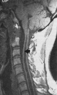
Magnetic resonance imaging has markedly improved our ability to study the anatomy and natural history of syrinx formation, but to date, our understanding of the process remains imprecise. This figure shows a longitudinal view of the spinal cord using MRI (magnetic resonance imaging). The arrow points to the syrinx which is seen to extend a great distance, from the cervical level down to the lower thoracic region. In general , the syrinx cavities are wider in the side to side dimension than in the AP dimension. In this T1 weighted image CSF is dark.