Longitudinal View
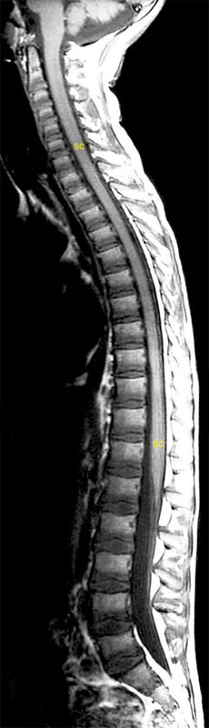
The spinal cord is seen using magnetic resonance imaging (MRI), in longitudinal view in the mid-sagittal plane. This is called a T1-weighted image, in which the CSF is dark.
The upper part includes the attachment to the brainstem, and the lower part includes the tapering conus medullaris, as well as the lumbar cistern.
Axial View
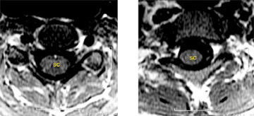
These two axial MRI views of the spinal cord show its size and location within the vertebral canal at two levels - C7 and T1 - these being the levels of the cord itself (not the vertebral levels). Again, the CSF is dark.