The Spinal Cord 'in situ'
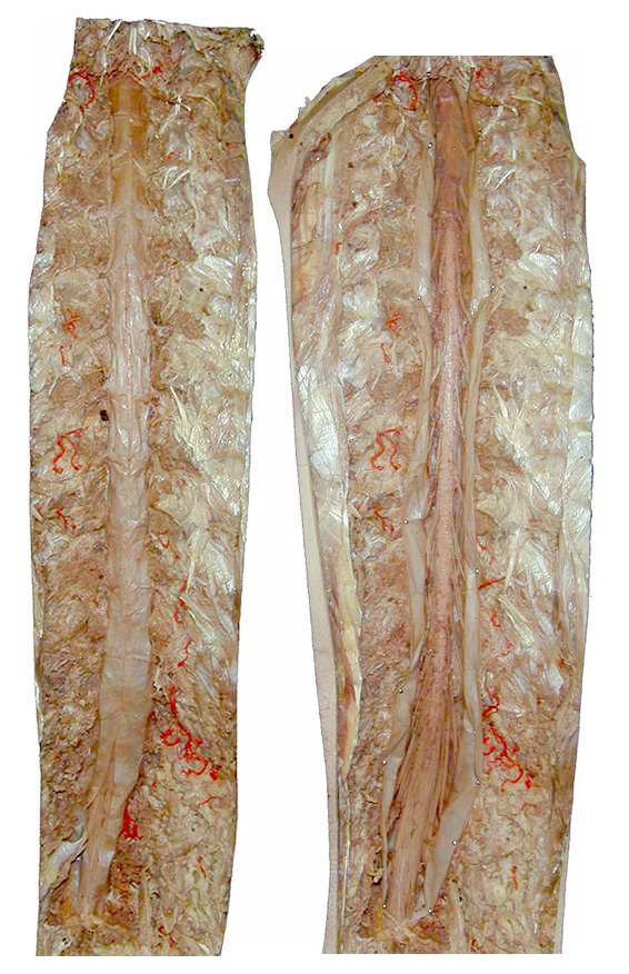
The spinal cord is shown 'in situ'in the vertebral (spinal) canal, from the posterior (dorsal) perspective, after removal of the muscles of the back and the vertebral arch of all the vertebrae.The dissected paraspinal muscles are seen, with some injected blood vessels, adjacent to the vertebral canal.
The view on the left shows the dura mater closed, and the view on the right shows the dura/arachnoid mater opened, allowing for visualization of the spinal cord.
The spinal cord ends at the level of the 2nd lumbar vertebra (in the adult) and the nerve roots contained within the lumbar cistern are collectively called the cauda equina [to be shown later in more detail].
Isolated Spinal Cord
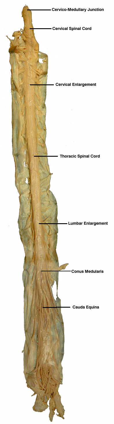
The isolated spinal cord is presented from a ventral (anterior) perspective. The attached sensory and motor nerve roots give the cord a 'segmented' appearance. Those nerves exiting/entering at the cervical level are almost horizontal, but as one descends to the thoracic and lumbar levels, the nerve roots orient in a more and more vertical manner, because they leave the spinal cord and must descend to the appropriate vertebral level to innervate their dermatome and myotome regions. The collection of nerve roots below the termination of the cord form the cauda equina which occupies the lumbar cistern [to be shown later in more detail].
NOTE that all nerve roots travel through the subarachnoid space, which is filled with CSF.
The spinal cord has two enlargements - cervical and lumbar (lumbosacral) - which mark the regions of the cord that innervate the upper and lower limbs respectively.
Cauda Equina
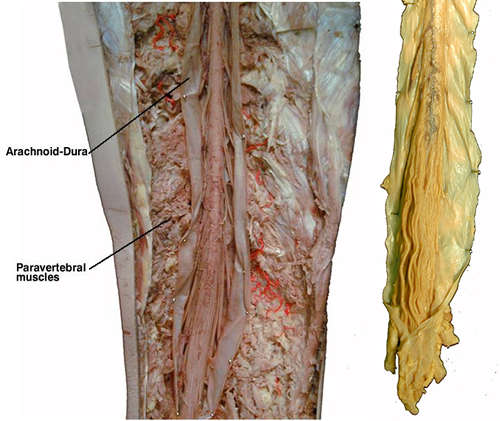
The nerve roots that form
the cauda equina are shown at higher magnification both 'in situ' and in the
isolated view of the spinal cord.