The mixed spinal nerves have both sensory (afferent) and motor (efferent) fibers, as well as sympathetic nerves.
The cell bodies for the sensory fibers are found in the dorsal root ganglia, located in the intervertebral spaces. The peripheral process of this neuron extends to the skin or muscles; the central process enters the spinal cord as the dorsal roots.
The motor nerves exit the spinal cord as the ventral roots, and join the sensory components beyond the dorsal root ganglion, thereby forming the mixed spinal nerve.
The cord is surrounded by meninges - pia, arachnoid (with the subarachnoid space containing cerebrospinal fluid), and dura.

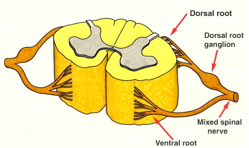
A representative segment
of the spinal cord is shown with attached nerves which are the roots:
The ventral or motor roots are exiting from the cord and going out to innervate
the muscles. The cell body for these nerves is the anterior (motor) horn cell
located in the ventral (anterior) horn of the spinal cord. A photographic view
of the ventral roots attached to the spinal cord is shown above.
NOTE: The denticulate ligaments are lateral extensions of the pia which attach to the arachnoid, presumably helping to 'stabilize' the spinal cord.
The dorsal or sensory roots are bringing in sensory information from the periphery (skin and muscles). The dorsal root ganglion (DRG), seen as an enlargement of the dorsal root, actually contains the cell bodies of these nerves. A photographic view of the dorsal roots attached to the spinal cord is shown above. Note that they are almost continuous. {The destination of these incoming sensory fibers is explained with the diagram <Sensory Fibers> next.}
The DRG is found as a swelling along the dorsal root - these enlargements can be seen in a view of the isolated spinal cord with attached nerve roots (see below). These DRG are actually located within the intervertebral spaces.
The mixed spinal nerve containing
motor and sensory (and also autonomic) fibers commences beyond (distal) to the
ganglion.